Thursday, February 27, 2014
Eyelid twitching "myokymia"
Flashes and Floaters
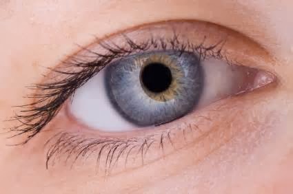
 Very common visual problems that people experience are flashes, floaters or a combination of each. To explain these issues in better detail, I first need to review eye anatomy.
The eye is a hollow structure filled with fluids. The front part of the eye has aqueous fliuid. It reside behind the cornea and in front of the colored iris tissue in a space called the anterior chamber. The majority of the eye is filled with a thick protein fluid called vitreous. It reside behind the iris in the vitreous cavity. Vitreous is like "raw egg whites." Lining the internal vitreous cavity is the retina vision tissue, which is like wallpaper.
Over time, the jello vitreous fluid begins to undergo chages - mostly shrinking and denaturaization. Small clumps of proten form. thse clumps cast a shadow obn the retina. Most apparent in bright light, agaist a plain background such as a white wall or blue sky. The floater shapes are varous: strings, strands, aomebas, bugs, nats, webs etc. These are usually benign but defintely pesky.
Floaters which are big, very dark grey, or block a large portion of your vision or occur with flashes ( "arcs / sparks" ) require immediate attention! Why? Well it may indicate a tear or detachment to the retinal tissue. The floaters here may be from a bleed or loose retinal tissue.
A dilated exam with a good search of the retina by a qualified eye doctor is the best course of action. If there is a small tear, a outpatient laser procedure can "spot weld" the loose retina back into place. A large tear or detachment may require major surgery.
For more information please visit www.myneweyes.com
Very common visual problems that people experience are flashes, floaters or a combination of each. To explain these issues in better detail, I first need to review eye anatomy.
The eye is a hollow structure filled with fluids. The front part of the eye has aqueous fliuid. It reside behind the cornea and in front of the colored iris tissue in a space called the anterior chamber. The majority of the eye is filled with a thick protein fluid called vitreous. It reside behind the iris in the vitreous cavity. Vitreous is like "raw egg whites." Lining the internal vitreous cavity is the retina vision tissue, which is like wallpaper.
Over time, the jello vitreous fluid begins to undergo chages - mostly shrinking and denaturaization. Small clumps of proten form. thse clumps cast a shadow obn the retina. Most apparent in bright light, agaist a plain background such as a white wall or blue sky. The floater shapes are varous: strings, strands, aomebas, bugs, nats, webs etc. These are usually benign but defintely pesky.
Floaters which are big, very dark grey, or block a large portion of your vision or occur with flashes ( "arcs / sparks" ) require immediate attention! Why? Well it may indicate a tear or detachment to the retinal tissue. The floaters here may be from a bleed or loose retinal tissue.
A dilated exam with a good search of the retina by a qualified eye doctor is the best course of action. If there is a small tear, a outpatient laser procedure can "spot weld" the loose retina back into place. A large tear or detachment may require major surgery.
For more information please visit www.myneweyes.com
Subscribe to:
Comments (Atom)